News Story
Real-time imaging platform for lab-on-CMOS biosensor systems

The proposed platform.
Complementary metal-oxide-semiconductor (CMOS)-based microelectronic sensors have great potential in the development of biosensors for medical and life science applications. Lab-on-CMOS microsystems incorporate one or more CMOS chips that can perform laboratory functions directly on the surface of the chips, allowing for intimate contact between sensing inputs and the cells under analysis. However, validating these lab-on-CMOS systems is a challenging task because of the difficulties in obtaining simultaneous ground-truth imaging and sensor data.
ISR-affiliated Professor Elisabeth Smela (ME), Professor Pamela Abshire (ECE/ISR), and two of Abshire’s graduate students, Bathiya Senevirathna and Sheung Luhave, have developed the first real-time imaging platform that generates high-quality images of lab-on-CMOS systems in cell culture environments. The platform was used to validate a CMOS capacitance sensor that monitors cell viability, proliferation, and death.
Several in vitro experiments were performed with human ovarian cancer cell lines where time-lapse images and capacitance recordings were performed simultaneously over three days. The images not only corroborated the temporal changes of capacitance recordings as the cells proliferated, but also unambiguously confirmed the sensor’s ability to detect single-cell binding events, track cell morphology changes, identify cell division events, and monitor cell motility.
“An Imaging Platform for Real-Time In Vitro Microscopic Imaging for Lab-on-CMOS Systems,” a paper based on their work, was presented at the 2019 IEEE Biomedical Circuits and Systems Conference (BioCAS).
Published December 11, 2019
Related Stories
Stories / July 9, 2021
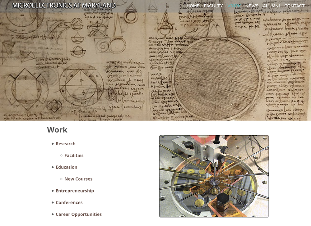
New website showcases work of Clark School’s microelectronics...
Stories / September 1, 2019
Alum Marc Dandin Appointed to Carnegie Mellon Faculty

Stories / June 19, 2017
Decade of TMV research leads to never-before-seen microsystems...
Stories / June 26, 2025
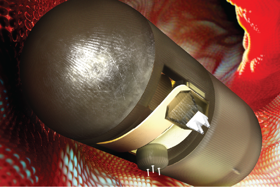
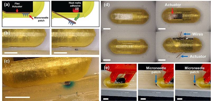
Adjustable Drug Release Marks New Milestone in Ingestible...
Stories / August 28, 2023
Miao Yu to develop cost-effective sensor for measuring lake...

Stories / July 28, 2023
Pneumatically controlled soft robotic catheters offer accuracy,...
Stories / May 21, 2023
New ‘FRRB’ packaging technology may solve an ingestible...
Stories / May 2, 2023
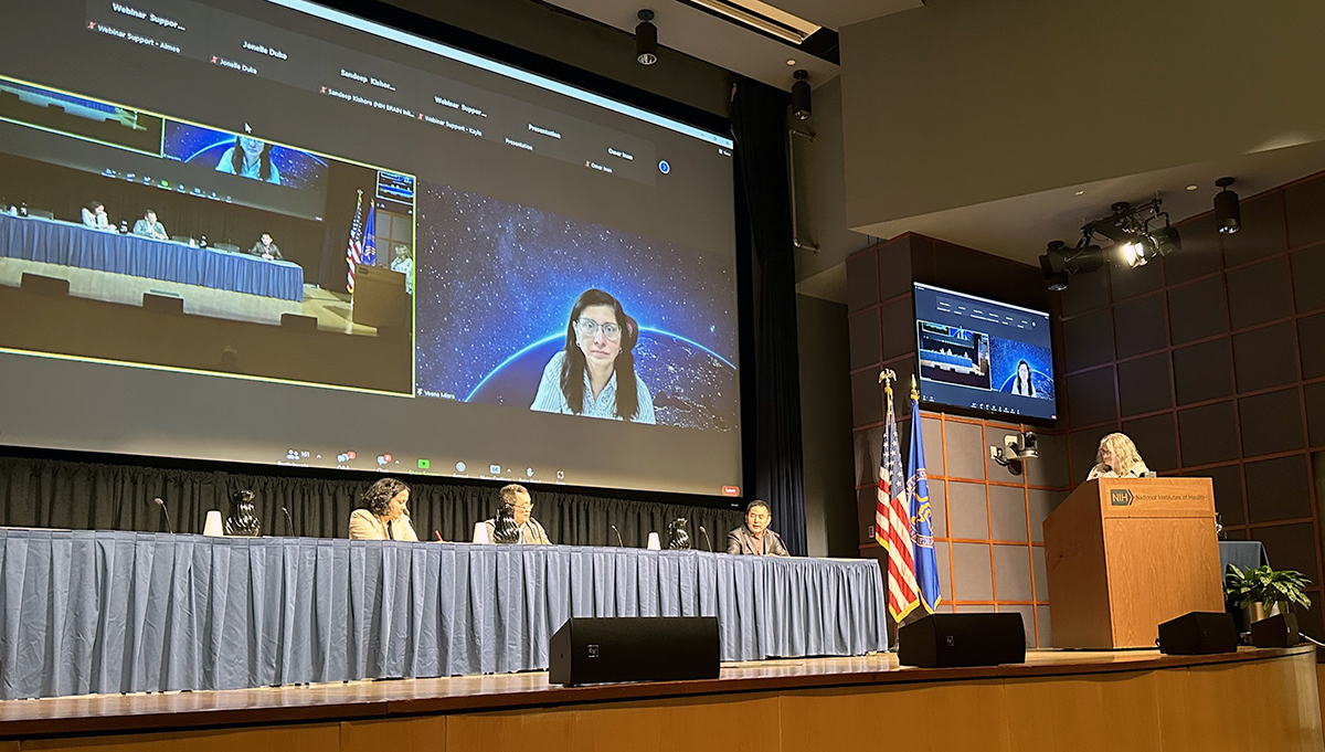
Ghodssi invited speaker at NIMH workshop on sensor technologies...
Stories / March 6, 2023
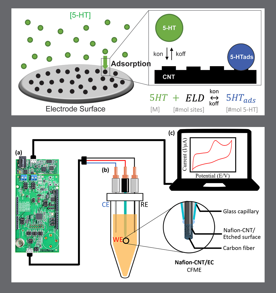
MSAL’s work on serotonin characterization and detection...
Stories / August 10, 2022
Undergrads and research experiences: Win! Win! Win!